Home
/ Animal Cell Under Electron Microscope Diagram : The Unit Of Life : V, w, x and are the organelles in the animal cell while structure z can be found in the nucleus.
Animal Cell Under Electron Microscope Diagram : The Unit Of Life : V, w, x and are the organelles in the animal cell while structure z can be found in the nucleus.
Animal Cell Under Electron Microscope Diagram : The Unit Of Life : V, w, x and are the organelles in the animal cell while structure z can be found in the nucleus.. In truth, there are still features of plant and animal cells we're only lately. Animal cell under electron microscope labelled. B) how the is structure labeled b adapted to its function? V, w, x and are the organelles in the animal cell while structure z can be found in the nucleus. The structure of cells be it a plant or animal cell is important because it forms the basic building block of the organism.
The complete guide for learning cell structure (with diagram). V, w, x dan y adalah organel dalam sel haiwan sementara struktur z boleh ditemui di The electron microscope electron microscopes use a beam of electrons instead of beams or rays of light. Maybe you would like to learn more about one of these?. Asked nov 28, 2017 in class.
Cell Upper Sec Science from joannewong.weebly.com Click (or tap) the diagram for a simple labelled version. To look at a cell close up we need a microscope. Thus, light microscopes allow one to visualize cells and their larger components such as nuclei, nucleoli, secretory granules, lysosomes, and large mitochondria. The electron microscope electron microscopes use a beam of electrons instead of beams or rays of light. A) name the parts labeled a and b. Depending on cellular function, one type of cell will have a higher number of certain organelles than others. Learn vocabulary, terms, and more with flashcards, games, and other study tools. Check spelling or type a new query.
You know, animal cell structure contains only 11 parts out of the 13 parts you saw in the plant cell diagram, because chloroplast and cell wall are available only in a plant cell.
Human cheek cell at 400x zoom. Below the basic structure is shown in the same animal cell, on the left viewed with the light microscope, and on the right with the transmission electron. For example, something that you draw as 3cm long, may in fact be 10, 000 times smaller in real life. Plant, animal and bacterial cells have smaller components each with the magnification of a microscope is not the only factor that is important when viewing. Cell structures as seen under the light and electron microscope cell structure under light microscope. Here's a diagram of a plant cell: Animal and plant cell under electron microscope. Examining plant cells under the microscope. Electron microscopy structure/function a cell contains organelles that are essential for its function. In blue, cell nuclei labelled with dapi. It uses a beam of electrons to illuminate the specimen instead of light as in the case of light microscope. That cells can be of different shapes and sizes. Plant cell under electron microscope labelled / what is a diagram of a plant and animal cell under an electron microscope quora / academia.edu is a platform for academics to share research papers.
Though we cannot see everything through the light microscope, some important organelles are visible and we can begin to see some structural differences. In the cell, there are organelles which are suspended within an aqeuos medium and contained. Asked nov 28, 2017 in class. Its this cell membrane that will contain all of the different parts of the cell. Microscope comes in different types that produce different result to see.
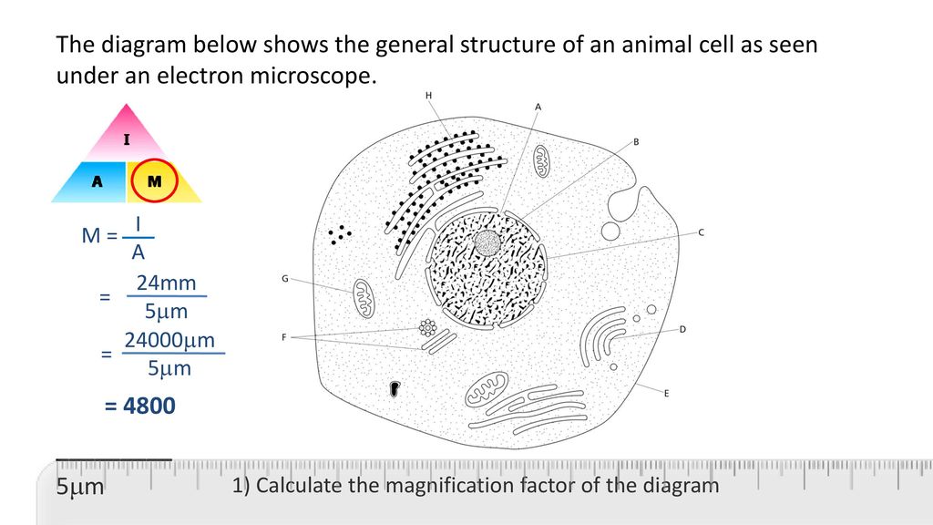
Microscopy And Magnification Ppt Download from slideplayer.com Microscope comes in different types that produce different result to see. Liver cell under electron microscope javesh bio2010 s weblog. The structure of cells be it a plant or animal cell is important because it forms the basic building block of the organism. Rajah 1.1 di bawah menunjukkan satu sel haiwan yang dilihat dibawah mikroskop elektron. The diagram below shows the general structure of an animal cell as seen under an electron microscope. In truth, there are still features of plant and anim. But at the same time it is interpretive. The structures within the cell are referred to as organelles.
Diagram 1.1 below shows an animal cell seen under the electron microscope.
1st john 1:1 holy hydrogen light of. To look at a cell close up we need a microscope. Examining plant cells under the microscope. But at the same time it is interpretive. Start studying plant cell under electron microscope. Maybe you would like to learn more about one of these? Below the basic structure is shown in the same animal cell, on the left viewed with the light microscope, and on the right with the transmission electron. Animal cells have a basic structure. Animal and plant cell under electron microscope. To check if you know the various cell organelles, examine the following schematic. Typical animal cell pinocytotic vesicle lysosome golgi vesicles golgi vesicles rough er (endoplasmic reticulum) smooth er (no ribosomes) cell (plasma) membrane mitochondrion golgi apparatus nucleolus nucleus centrioles (2) each composed of 9 microtubule triplets microtubules cytoplasm ribosome The structure of cells be it a plant or animal cell is important because it forms the basic building block of the organism. Click (or tap) the diagram for a simple labelled version.
Here's a photo of a plant cell under an electron microscope. Illustrate only a plant cell as seen under electron microscope how. V, w, x and are the organelles in the animal cell while structure z can be found in the nucleus. A) name the parts labeled a and b. The figure below is a fine structure of a generalized animal cell as seen under an electron microscope.
Electron Microscopic Study Of Cell And Organelles Important from i2.wp.com Diagram 1.1 below shows an animal cell seen under the electron microscope. Prokaryotic cell under light microscope tescar. Under the microscope, animal cells appear different based on the type of the cell. Electron microscope can magnify an object up to 500, 000 times. Animal cells have a basic structure. Its this cell membrane that will contain all of the different parts of the cell. Be sure you follow all the correct procedures when using the microscope to avoid damage to your equipment, your samples, and yourself. The structures within the cell are referred to as organelles.
Here's a diagram of a plant cell:
The diagram is very clear, and labeled; They are very tiny than what human eyes can see in general. The structure of cells be it a plant or animal cell is important because it forms the basic building block of the organism. Electron microscope can magnify an object up to 500, 000 times. Here's a photo of a plant cell under an electron microscope. Liver cell under electron microscope javesh bio2010 s weblog. In truth, there are still features of plant and animal cells we're only lately. Plant cell under electron microscope labelled / what is a diagram of a plant and animal cell under an electron microscope quora / academia.edu is a platform for academics to share research papers. V, w, x and are the organelles in the animal cell while structure z can be found in the nucleus. Living cells cannot be observed using an electron microscope because samples are placed in a. Maybe you would like to learn more about one of these?. Even at very high magnification, they look like (pairs of) dots. Rajah 1.1 di bawah menunjukkan satu sel haiwan yang dilihat dibawah mikroskop elektron.
Share :
Post a Comment
for "Animal Cell Under Electron Microscope Diagram : The Unit Of Life : V, w, x and are the organelles in the animal cell while structure z can be found in the nucleus."


Post a Comment for "Animal Cell Under Electron Microscope Diagram : The Unit Of Life : V, w, x and are the organelles in the animal cell while structure z can be found in the nucleus."