Home
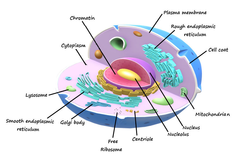
/ Draw And Label A Neat Diagram Of Animal Cell - Draw A Diagram Of Typical Cell And Label The Following Parts In It Cell Membranevacuolenucleusendoplasmic Reticulummitochondriagolgi Body : Under the microscope, an animal cell shows many different parts called organelles, that work together to keep the cell functional.
Draw And Label A Neat Diagram Of Animal Cell - Draw A Diagram Of Typical Cell And Label The Following Parts In It Cell Membranevacuolenucleusendoplasmic Reticulummitochondriagolgi Body : Under the microscope, an animal cell shows many different parts called organelles, that work together to keep the cell functional.
Draw And Label A Neat Diagram Of Animal Cell - Draw A Diagram Of Typical Cell And Label The Following Parts In It Cell Membranevacuolenucleusendoplasmic Reticulummitochondriagolgi Body : Under the microscope, an animal cell shows many different parts called organelles, that work together to keep the cell functional.. Draw a neat labelled diagram of an animal cell. The cell membrane of an animal cell is not a perfect most instructors will make you to label each structure on a test or assignment. Under the microscope, an animal cell shows many different parts called organelles, that work together to keep the cell functional. Label the animal cell diagram using the attached glossary of animal cell terms. Draw a neat diagram of plant cell and label any three parts which differentiate it from animal cell.
Asked feb 5, 2018 in class ix science by saurav24 expert (1.4k points). Though this animal cell diagram is not representative of any one particular type of cell it provides insight into the primary organelles and the intricate internal draw a neat diagram of plant cell and label any three parts which image information: Some cells have specialised functions. As observed in the labeled animal cell diagram the cell membrane forms the confining factor of the cell that is it. Cell membranes do allow molecules to pass in and out of animal cells.
Draw A Neat Diagram Of A Animal Cell B Plant Cell C Algea Cell D Bacteria E Paramissium Science Cell Structure And Functions 12967751 Meritnation Com from www.winkelhage.com Draw a neat diagram of plant cell and label any three parts which differentiate it from animal cell. In fact, most are invisible without using a microscope. Those are the main parts of a cell in an animal that you will have to draw. How to draw a animal cell easy and step by step. Printable animal cell diagram to help you learn the organelles in an animal cell in preparation for your test or quiz. However, in plant cells, centrioles or centrosomes are absent, but still microtubule formation takes place through mitotic phases of cell division. Using arrows and textables, label each part of the cell and describe its function. In the labeled animal cell diagram, it is nearly circular in shape and lacks outer cell wall;
Label the animal cell diagram using the attached glossary of animal cell terms.
As observed in the labeled animal cell diagram the cell membrane forms the confining factor of the cell that is it. With a wider definition, lysosomes are found in the cytoplasm of plant and protists as well as animal cell. The cell membrane of an animal cell is not a perfect most instructors will make you to label each structure on a test or assignment. Include descriptions of what each part does. Animal cell structure animal cells are typical of the eukaryotic cell enclosed by a plasma membrane and containing a membrane bound nucleus and organelles. Let`s draw a typical animal cell. How to draw a animal cell easy and step by step. Draw a neat diagram of plant cell and label any three parts which differentiate it from animal cell. The parts of an animal cell have distinct functions. Find diagrams of a plant and an animal cell in the science tab. • a eukaryotic cell divides once in every 24hrs. Draw a neat labelled diagram of an animal cell. Cells form the basic building blocks for all living things.
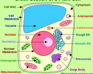
Find diagrams of a plant and an animal cell in the science tab. Let`s draw a typical animal cell. Draw a neat diagram of the stomatal apparatus found in the epidermis of leaves and label the stoma, guard cells, chloroplast, epidermal cells, cell. Draw a neat diagram of plant cell and label any three parts which differentiate it from animal cell. 2.3.1 draw and label a diagram of the ultrastructure of a liver cell as an example of an animal cell.
Draw A Neat Labeled Diagram Of An Animal Cell Class 11 Biology Cbse from www.vedantu.com • a eukaryotic cell divides once in every 24hrs. Animal cells contain organelles known as centrioles which are not present in plant cells. 2.3.1 draw and label a diagram of the ultrastructure of a liver cell as an example of an animal cell. How to draw the animal cell diagram 4. How to draw a animal cell easy and step by step. Printable animal cell diagram to help you learn the organelles in an animal cell in preparation for your test or quiz. In the labeled animal cell diagram, it is nearly circular in shape and lacks outer cell wall; 17 010 просмотров • 10 авг.
The fundamental unit of life.
• interphase (22hrs approx.) this is the resting phase of. Draw a neat labelled diagram of an animal cell. Draw a neat diagram of plant cell and label any three parts which differentiate it from animal cell. Though this animal cell diagram is not representative of any one particular type of cell it provides insight into the primary organelles and the intricate internal draw a neat diagram of plant cell and label any three parts which image information: All of tims printables in one draw this animal cell by following this drawing lesson. The cell is the basic unit of life. Animal cells have a single highly complex and prominent golgi apparatus. The animal cells perform different roles in the human body, which play a crucial role in those bodily functions. As observed in the labeled animal cell diagram the cell membrane forms the confining factor of the cell that is it. Draw a neat diagram of plant cell and label any three parts which differentiate it from animal cell. Find diagrams of a plant and an animal cell in the science tab. Include descriptions of what each part does. Animal cells contain organelles known as centrioles which are not present in plant cells.
Then, label this the plasma membrane. While the plant cell resembles rectangular shape and. 17 010 просмотров • 10 авг. In the above diagram there is a dry cell with description of its parts. Under the microscope, an animal cell shows many different parts called organelles, that work together to keep the cell functional.
Structure Of Cell Cell Structure And Functions Class 8 from classnotes.org.in Printable animal cell diagram to help you learn the organelles in an animal cell in preparation for your test or quiz. Drawing cells is typically not a skill assessed on tests or required by standards, but it can certainly help students develop a lasting knowledge of the cell. Draw a neat labelled diagram of an animal cell. Asked feb 5, 2018 in class ix science by saurav24 expert (1.4k points). Then, label this the plasma membrane. Some cells have specialised functions. Cell membranes do allow molecules to pass in and out of animal cells. 461 x 503 pixel type jpg download.
Draw a neat diagram of plant cell and label any three parts which differentiate it from animal cell.
A lysosome is a cell organelle. Draw a neat labelled diagram of an animal cell. Printable animal cell diagram to help you learn the organelles in an animal cell in preparation for your test or quiz. Animal cells have a single highly complex and prominent golgi apparatus. Vacuoles in animal cells are many and small. 2.3.2 annotate the diagram from 2.3.1 with the functions of each named structure. Most cells are very small; Label both a plant and animal cell on a poster layout. Animal cells differ from plant cells in several regards though, including the lack of vacuoles draw a simple circle or oval for the cell membrane. How to draw the animal cell diagram 4. Get free solutions to all questions from chapter the fundamental unit of life. Since animal cells are softer than plant cells centrioles are required to. Under the microscope, an animal cell shows many different parts called organelles, that work together to keep the cell functional.
Share :
Post a Comment
for "Draw And Label A Neat Diagram Of Animal Cell - Draw A Diagram Of Typical Cell And Label The Following Parts In It Cell Membranevacuolenucleusendoplasmic Reticulummitochondriagolgi Body : Under the microscope, an animal cell shows many different parts called organelles, that work together to keep the cell functional."


Post a Comment for "Draw And Label A Neat Diagram Of Animal Cell - Draw A Diagram Of Typical Cell And Label The Following Parts In It Cell Membranevacuolenucleusendoplasmic Reticulummitochondriagolgi Body : Under the microscope, an animal cell shows many different parts called organelles, that work together to keep the cell functional."